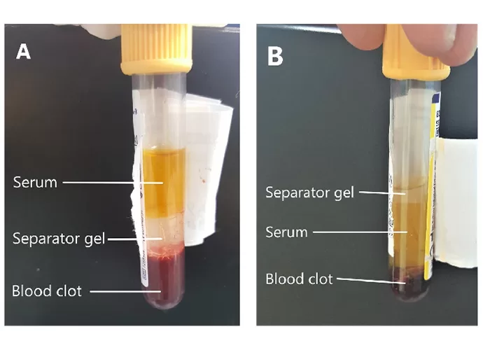
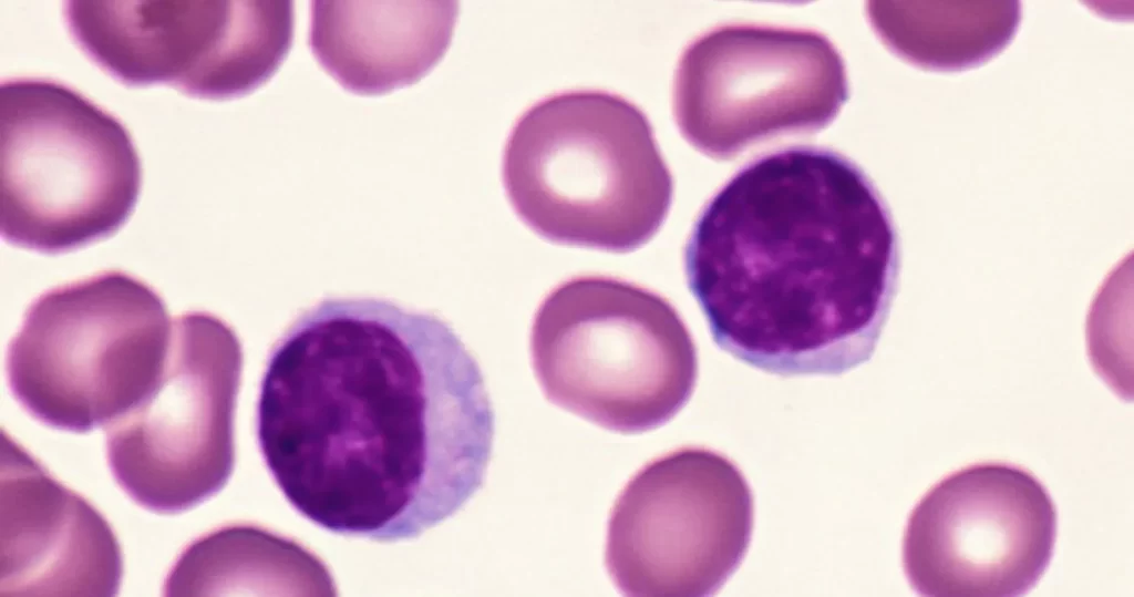
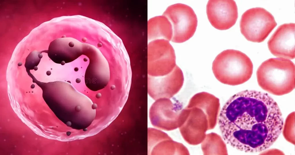
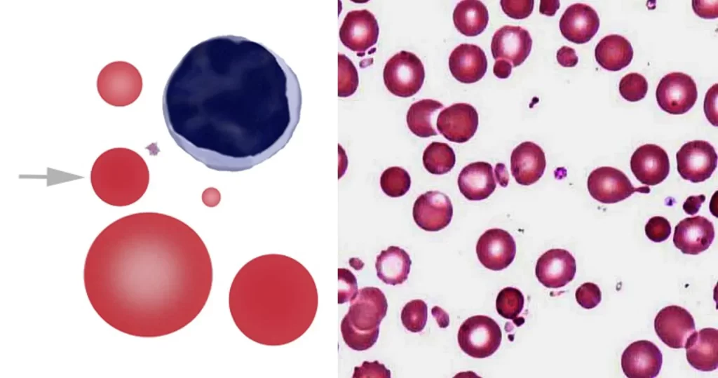
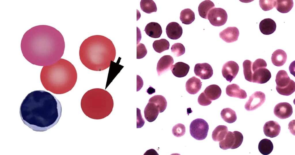
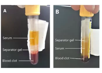
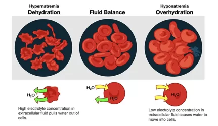
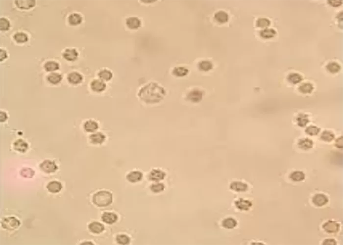
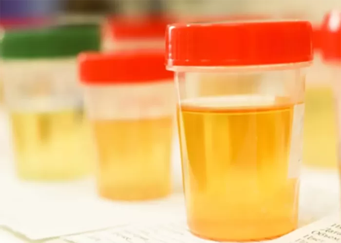
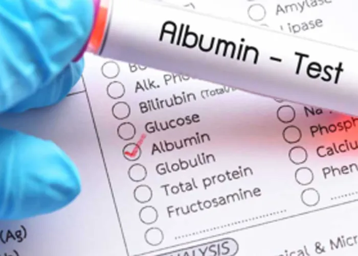
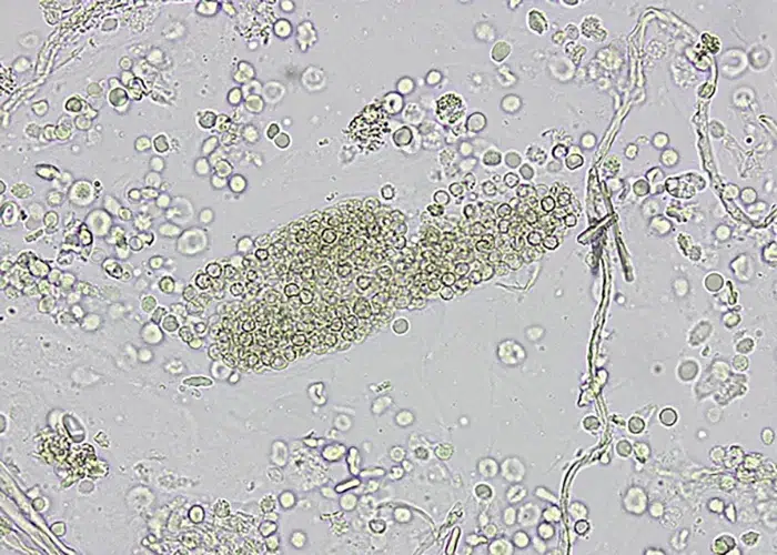
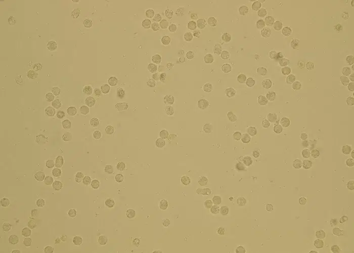
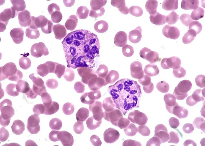
In live blood analysis, serum refers to the liquid portion of the blood that remains after the blood cells have been separated through clotting. It is a key focus in non-magnified blood observations because it provides valuable information about the biochemical state of the body. The serum contains proteins such as albumin and globulins, electrolytes like sodium and potassium, as well as metabolic waste products and hormones. Observing the serum in its natural state can help identify imbalances, nutrient deficiencies, or the presence of inflammatory markers.